At a Glance
• The most important characteristic of a vital dye is its ability to differentiate among intraocular tissues to allow delineation and removal of target membranes.
• The ideal posterior segment dye would address all intraocular tissue structures of relevance in vitreoretinal surgery.
• Use of a combination dye can simplify chromovitrectomy staining overall.
The Ideal Dye for Posterior Staining
By Andreas M. Mohr, MD
In selecting a vital dye for posterior staining, the most important characteristic that vitreoretinal surgeons look for is the ability to differentiate among thin, translucent intraocular tissues to allow delineation and removal of target membranes. In the past, surgeons may have focused on identifying and removing the epiretinal membrane (ERM), but other tissues such as the internal limiting membrane (ILM) and posterior hyaloid have become bigger focuses with regard to obtaining good final visual outcomes in certain macular diseases, as these membranes may coexist in certain pathologies. All of these key intraocular structures must be visually differentiated with an effective vital staining procedure. This article reviews the characteristics of commonly used dyes, describes the qualities that an ideal dye for use in vitreoretinal surgeries would possess, and examines the benefits of combination dyes.
THE USUAL SUSPECTS
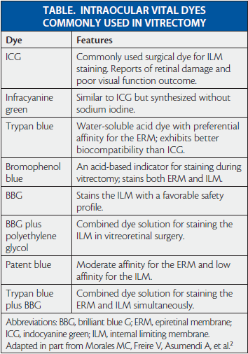
Trypan blue; 0.025% brilliant blue G (BBG) plus 4% polyethylene glycol (PEG; ILM-Blue, DORC International); and indocyanine green (ICG; IC-Green, Akorn) are the dye preparations most often used for posterior staining (Table).
Trypan Blue
Trypan blue has a high affinity for the ERM, whereas BBG and other dye formulations focus more on the ILM. Trypan blue is a useful staining agent in vitreoretinal surgery, but it has toxic effects if left in the eye for long periods (> 30 minutes).1 Purification to remove potential contaminants arising from the production process helps to maximize its safety.
Indocyanine Green
Although ICG has a good staining effect in vitreoretinalsurgery, there are phototoxicity concerns associated with certain concentrations, staining time, duration on the posterior pole, and osmolarity.2,3 ICG should be considered for use only by experienced surgeons; however, its staining effect may be useful for beginners.
Brilliant Blue G
BBG is more translucent than trypan blue.4 The latter, depending on the manufacturer, can have a purity level between 80% and 97%. Impurities can cause serious inflammation in the eye membranes.
A study by Williamson and Lee evaluating visual outcomes of idiopathic macular hole surgeries using different staining solutions found that better outcomes were achieved with BBG (0.25 mg/L) for ILM peel than with ICG (0.5 mg/mL).5
A multicenter clinical assessment of two high molecular weight dyes—BBG and the combination of 0.15% trypan blue, 0.025% BBG, and 4% PEG (MembraneBlue-Dual, DORC International)—for membrane staining during macular surgery showed that either dye can be injected into a fluid-filled vitreous cavity to facilitate staining and removal of the ILM and ERM without additional air-fluid exchange.6,7
Characteristics of the Ideal Liquid Staining Solution for Vitreoretinal Surgery
• Enables consistent identification and visualization throughout the delayering or peeling procedure
• Easy to use for experienced and inexperienced surgeons
• Provides a viscous and dense solution to avoid unwanted dispersion
• Allows simultaneous staining of the ERM and ILM without additional air-fluid exchange
• Has minimal toxicity or safety concerns
• Yields enhanced color-staining contrast
THE IDEAL DYE
The ideal posterior segment dye would address all intraocular tissue structures of relevance in vitreoretinal surgery, allowing consistent identification and visualization of tissue layers throughout the delayering or peeling procedure. It would be ready to use not only for beginners who may need more time for tissue identification during vitreoretinal surgery but also for experienced surgeons. Another key feature of the ideal posterior staining solution would be the ability to adequately delineate different ocular structures (see “Characteristics of the Ideal Liquid Staining Solution for Vitreoretinal Surgery”).
COMBINED USE OF DYES
Assessments using thin layer chromatography have shown that combining trypan blue and BBG did not result in a consolidated staining effect.1 The two dyes did not interact chemically with one another; rather, each dye maintained differential structures, demonstrating that trypan blue has a completely different chemical action from that of BBG.
A combined solution that contains trypan blue, BBG, and PEG offers a more viscous and denser solution. Additionally, in vitro studies evaluating the influence of combination dyes on cell damage demonstrate that BBG protects against trypan blue toxicity, while a combination of trypan blue, BBG, and PEG is less toxic to retinal pigment epithelial cells than trypan blue alone.1
CONCLUSION
In my surgical experience, using a combination dye simplifies chromovitrectomy staining overall. In at least 50% of cases, when you remove the ERM you partially remove some of the ILM, but it is difficult to identify which part has been removed using trypan blue alone. With a dual dye that contains BBG, you achieve good visualization throughout the vitreous surgery procedure and avoid the need for an additional fluid-air exchange. For these reasons, combination staining is the future for simultaneous staining of the ERM and ILM.
Developments in ophthalmic vitreoretinal liquids are likely to focus on additional vital dye agents that offer enhanced color contrast against an orange-red retina, perhaps with a more greenish tint for maximum visible contrast. n
Andreas M. Mohr, MD, is a member of the clinical competence center at the University of Bremen and is head of the Eye Hospital, St. Joseph Stift, Bremen, Germany. He has financial relationships with Alcon, Novartis, Bayer Healthcare, Allergan, and DORC. Dr. Mohr may be reached at dramohr@yahoo.de.
1. Awad D, Schrader I, Bartok M, et al. Comparative toxicology of trypan blue, brilliant blue G, and their combination together with polyethylene glycol on human pigment epithelial cells. Invest Ophthalmol Vis Sci. 2011;52(7):4085-4090.
2. Morales MC, Freire V, Asumendi A, et al. Comparative effects of six intraocular vital dyes on retinal pigment epithelial cells. Invest Ophthalmol Vis Sci. 2010;51(11):6018-6029.
3. Creuzot-Garcher C, Acar N, Passemard M, et al. Functional and structural effect of intravitreal indocyanine green, triamcinolone acetonide, trypan blue, and brilliant blue G on rat retina. Retina. 2010;30(8):1294-1301.
4. Wu L, Velasquez R, Montoya O. Non-infectious endophthalmitis associated with trypan blue use in cataract surgery. Int Ophthalmol. 2008;28(2):89-93.
5. Williamson TH, Lee E. Idiopathic macular hole: analysis of visual outcomes and the use of indocyanine green or brilliant blue for internal limiting membrane peel. Graefes Arch Clin Exp Ophthalmol. 2014;252(3):395-400.
6. Veckeneer M, Mohr A, Alharthi E, et al. Novel ‘heavy’ dyes for retinal membrane staining during macular surgery: multicenter clinical assessment. Acta Ophthalmol. 2014;92(4):339-344.
7. Mohr A, Bruinsma M, Oellerich S, et al; International Chromovitrectomy Collaboration. Dyes for eyes: hydrodynamics, biocompatibility and efficacy of ‘heavy’ (dual) dyes for chromovitrectomy. Ophthalmologica. 2013;230(suppl 2):51-58.


_1784132761.jpg?auto=compress,format&w=75)


