A 61-year-old man presented with a sudden reduction in vision in his right eye (OD) lasting 3 days. His BCVA at presentation was 20/200 in each eye (OU). He had a history of cataract surgery OD performed 4 years earlier. Slit-lamp examination showed a normal anterior segment with pseudophakia OD and advanced cataract in the patient’s left eye (OS). Fundus examination revealed a complete retinal detachment OD with a giant retinal tear that extended from the 7 o’clock position to the 11 o’clock position, another small retinal tear at 3 o’clock, and a full thickness macular hole (Main Figure).
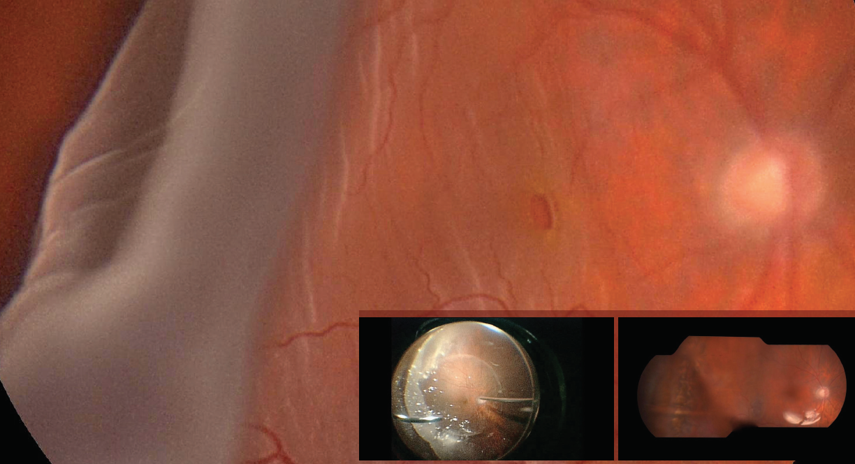
Vitrectomy was performed using 23-gauge instrumentation. The edges of the tear were unrolled, and complete retinal reattachment was achieved under perfluorocarbon liquid. Intraoperative endolaser was performed around the peripheral retina for 360° and around the edges of the tears. Perfluorocarbon liquid was exchanged with silicone oil 5000 cs as final tamponade (Inset, left).
At the 6-month follow-up visit, the patient’s retina was attached and the macular hole was repaired. It is possible to appreciate the wide scar in the temporal quadrant as a consequence of the giant retinal tear and the surrounding laser photocoagulation (Inset, right). A mild rise in intraocular pressure was successfully managed with a combination topical beta blocker and carbonic anhydrase inhibitor. Silicone oil tamponade remains in situ and is scheduled to be removed 9 months after the date of the primary vitrectomy. The patient’s BCVA is 20/125 OD.
Section Editor Manish Nagpal, MS, DO, FRCS (Edin)
• Senior Consultant, Retina and Vitreous Services, the Retina Foundation, Ahmedabad, India
• drmanishnagpal@yahoo.com
• Financial disclosure: None
Matteo Forlini, MD
• Vitreoretinal Specialist, Domus Nova Hospital, Ravenna, Italy
• matteoforlini@gmail.com
• Financial disclosure: None
Alessandro Romani, MD
• Ophthalmology Resident, Institute of Ophthalmology, University of Parma, Parma, Italy
• alessandro.romani1@studenti.unipr.it
• Financial disclosure: None
Purva Date, DNB, FVRS
• Vitreoretinal Specialist, Aditya Jyot Eye Hospital, Wadala, Mumbai, India
• drpurvadate@yahoo.in
• Financial disclosure: None
If you have an image or images you would like to share, email Dr. Nagpal.
Note: Photos should be 400 dpi or higher and at least 10 inches wide.


_1784132761.jpg?auto=compress,format&w=75)


