
A 15-year-old child presented to us after a cricket ball injury to his left eye with multiple large retinal tears. These tears were located in the inferior and temporal quadrants extending across 2 to 2.5 clock hours circumferentially (Figure1). Unlike a dialysis, these tears had anterior flaps and extended quite posteriorly up to the equator (Figure 1). The tears were irregular and had ragged edges with fresh retinal and preretinal hemorrhages (Figure 2). The tears were also surrounded by a cuff of subretinal fluid that merged into the areas of commotio retinae (Figures 1 and 2).
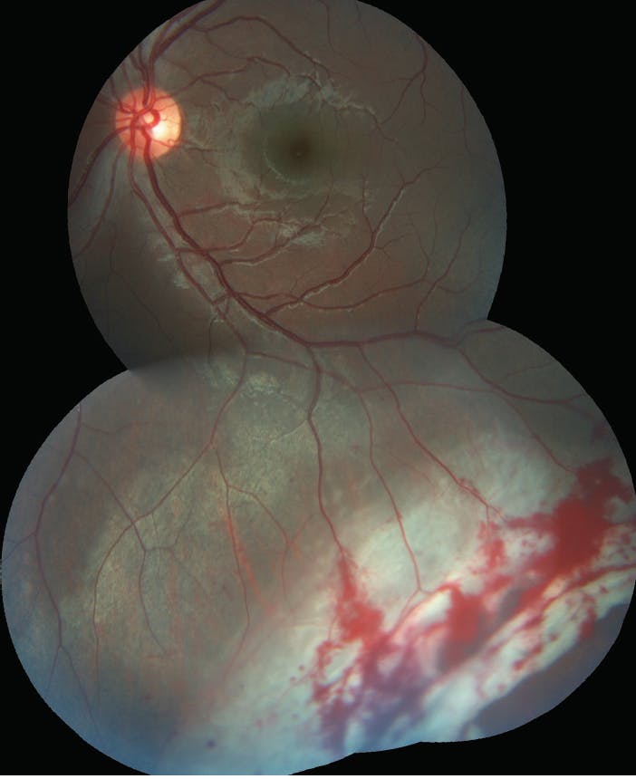
Figure 1. Retinal tears with subretinal fluid and commotio retinae located in the inferior and temporal quadrant extending up to the equator.
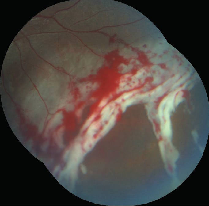
Figure 2. Close-up image showing the irregular and ragged tears with fresh retinal and preretinal hemorrhages.
The VA was 20/20 OS. The child did not complain of any defect in his visual field. After weighing the risks and benefits of various treatment options, we decided to perform an aggressive delimitation of the entire lesion with four to five contiguous rows of laser around the tears and subretinal fluid posteriorly, extending up to the ora serrata anteriorly (Figure 3). We chose laser delimitation as it represented the least invasive treatment option, compared to a large buckle or extensive vitreous surgery with tamponade.
The child continues do well with 20/20 VA OS, with no evidence of progression of subretinal fluid for over a year following the treatment.

-1_1607102916.jpg?auto=compress,format&w=70)


_1784132761.jpg?auto=compress,format&w=75)


