
In March 2020, our world turned upside down. Until then, everyone, including retina specialists, was basking in the luxury of the predictable. We scheduled our patients as often as we considered necessary. To achieve good visual results, we knew we needed to follow meticulous monitoring and treatment routines, and so we did.
But with the COVID-19 pandemic, it became abundantly clear to the ophthalmology community that things needed to change. At first, we believed the upheaval would be short-lived, so we treated only the urgent patients. In some countries and institutes, this meant seeing patients with AMD and choroidal neovascularization who were being treated with anti-VEGF agents, but rescheduling patients with diabetic macular edema (DME), assuming the persistent edema in the latter group could wait a few weeks or months until the pandemic was over. We treated urgent cases of retinal detachment, of course, but did not perform elective cataract surgeries.
Unfortunately, the pandemic has lasted well beyond those first weeks and months, and we could not continue to postpone treatment for our nonurgent patients. We started to witness severe irreversible decreases in vision due to the lack of treatment from missed appointments.1 Finding a way to treat all of our patients in the midst of the pandemic became our highest priority. The solution my department decided to adopt was to decentralize ophthalmic care based on a four-part strategy.
PART 1: ENSURING A SAFE ENVIRONMENT
First, we had to provide patients a safe and protective space by taking all necessary precautions and measures against COVID-19. We prescreened patients to ensure that none were experiencing fever or flu-like symptoms. Protective face masks and social distancing on the patients’ part went without saying, and the latter was achieved by moving furniture and roping off chairs in our waiting rooms.
We also provided personal protective equipment for our staff. In addition to wearing a face mask, our ophthalmologists sit behind a plastic shield that separates them from the patient during slit-lamp evaluations, laser treatments, and imaging studies (Figure 1).
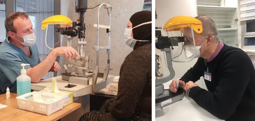
Figure 1. To reduce the risk of exposure, the ophthalmologist sits behind a protective shield when examinations require close contact with a patient.
We also reduced the patient load by increasing our clinic hours. We called patients the day before their scheduled appointments to ensure that they planned to come, and we did our best to convince reluctant patients to keep their appointments. Those who did not wish to attend were immediately rescheduled to avoid a future backlog.
Another aspect of ensuring a safe treatment environment was to reduce the length of appointments as much as possible. Our physicians began reviewing the patient’s charts and imaging studies (accessible via the institution’s electronic medical records database) before summoning the patient to the consultation room. The examination could then be carried out as soon as the patient was seated.
Some ophthalmologists suggested skipping visual acuity evaluations at each visit. To explore the ramifications of such a change, I conducted a small study to assess whether adding a visual acuity finding to the electronically available data led to significant changes in decision-making. The results revealed that visual acuity evaluations caused a change in management less than 10% of the time. We accepted that as a reasonable level of risk compared with the considerable gain in expediency.
We also considered other changes that could save time and yet carried no risk to patients with AMD: that is, switching patients to a longer-acting drug and changing their regimen to either a treat-and-extend or to a fixed regimen. With these regimens, we saw patients only on the day of injection rather than scheduling monitoring appointments.
PART 2: SEPARATING RETINA CARE
We also endeavored to isolate the ophthalmology visit from the high-risk hospital environment. We opened a clinic in a remote part of the city that allowed us to provide shorter waiting times and a more spacious setting (Figure 2). The office space—a generous donation of WeWork Israel—became available when many of their offices were vacated when people began working from home due to the COVID-19 pandemic.

PART 3: EMBRACING TELEMEDICINE
We also decided to establish an integrative telemedicine clinic. At each office visit, the physician evaluated the patient’s suitability for a telemedicine visit for the next follow-up. This was especially useful for patients with external eye disease who could photograph themselves and send an image during the virtual visit. It was also helpful for patients who underwent OCT or other imaging studies at another location.
Importantly, we made arrangements at the hospital management level with the HMO payers to ensure that these visits were reimbursed.
PART 4: INSTITUTING IN-HOME CARE
Our most innovative step was establishing outreach to individual patients’ homes. The mission was to treat those patients with little or no access to health care, as well as patients who were too fragile or ill to leave their homes during the pandemic, all with the aim of preventing deterioration in vision.
Our patient selection strategy was meticulous: We located the patients who missed appointments, were very elderly, had systemic risk factors, or whose disease was one of medical priority (eg, choroidal neovascularization secondary to AMD as opposed to DME). We established a dedicated communication team that evaluated patients for these parameters.
After checking that the home setting was not a risk factor for endophthalmitis, we organized a mobile unit in which the ophthalmologist and a nurse or technician traveled to the patient’s home and carried out the necessary injections (Figures 3 and 4).


Figure 4. With the right tools and training, ophthalmologists can perform in-home injections for the most high-risk patients.
No published studies have compared the incidence of infection after intravitreal injection in patients’ homes versus the clinic setting, although a few studies have compared the impact of carrying out intravitreal injections in the OR compared with the clinic. Those studies concentrated on the incidence of endophthalmitis and, for the most part, showed no difference between locations.2 As a point of clarification, most intravitreal injections in Israel are done in the clinic, not in a sterile environment.
THE RIGHT TOOLS FOR THE JOB
The unprecedented adjustments we have made highlight the benefits of many emerging treatment options. For example, we now have drugs with a longer duration of action. The FDA-approved anti-VEGF agent brolucizumab-dbll (Beovu, Novartis) can be administered every 3 months, although it remains under investigation regarding the risk of developing intraocular inflammation. Another promising option is faricimab (Roche), an inhibitor of VEGF and angiopoietin-2, which has met the primary endpoint in two phase 3 studies in DME; it is currently under regulatory consideration by the FDA and, if approved, can be administered every 3 or potentially even every 5 months.
An especially exciting development is the Port Delivery System (Roche). The phase 3 Archway trial demonstrated that a refill every 6 months was not inferior in efficacy to monthly ranibizumab (Lucentis, Genentech) injections in terms of improvement in visual acuity and reduction in central retinal thickness.
We look forward to adopting these longer-duration drugs and adding them to our armamentarium of treatments for patients with AMD or DME. However, these longer-acting drugs will require superior techniques for monitoring therapeutic response. Not all of our patients (albeit more than 50%) will be able to benefit from the longer intervals between injections.
Another exciting innovation is the development of at-home OCT (Notal Home OCT, Notal Vision), which has been evaluated in a few studies with successful imaging achieved in 93% of the enrolled eyes.3 Positive and negative agreement for detection of fluid, intraretinal fluid, and subretinal fluid in at least one of three consecutive spectral-domain OCT images was 97%/95%, 96%/94%, and 100%/98%, respectively, with the Notal Home OCT compared with commercial in-office OCT systems. As many as 95% of patients reported that it was easy to operate the device without assistance.
The analysis and depiction of fluid distribution and volume in a longitudinal case study of the Notal Home OCT illustrated the acute nature of wet AMD and the therapeutic response to anti-VEGF injections. The researchers concluded that the at-home OCT system met the requirements for self-controlled imaging by wet AMD patients with regard to image quality, field of view, and usability.
The expectation is that image analysis based on artificial intelligence can potentially support clinicians in the assessment and use of large amounts of data generated by daily at-home OCT imaging.3
KEY TAKEAWAYS
To provide the excellence in ophthalmic care to which we are committed, even in times of pandemic, we must be flexible. We must adapt to new situations, innovate, think outside the box, and dare to try something new.
How will the field of ophthalmology look after the COVID-19 pandemic is finally behind us? I believe our herculean and often exhausting efforts will bring ongoing changes to the ways we provide exceptional care to our patients—pandemic or not.
1. Romano F, Monteduro D, Airaldi M, et al. Increased number of submacular hemorrhages as a consequence of coronavirus disease. Ophthalmol Retina. 2020;4(12):1209-1210.
2. Tabandeh H, Boscia F, Sborgia A, et al. Endophthalmitis associated with intravitreal injections: Office-based setting and operating room setting. Retina. 2014;34(1):18-23.
3. Nahen K, Beniamini G, Loewenstein A. Evaluation of a self-imaging SD‐OCT system for remote monitoring of patients with neovascular age related macular degeneration. Klin Monatsbl Augenheilkd. 2020;237:1410-1419.


_1784132761.jpg?auto=compress,format&w=75)


