

A 96-year-old woman with history of vision loss OD and gradually worsening vision OS presented with BCVA OD CF at 1 ft and BCVA OS 20/60.
Examination OD revealed neovascular age-related macular degeneration with drusen, RPE tear, fibrovascular choroidal neovascular membrane, and subretinal hemorrhage. The patient underwent therapy with bevacizumab (Avastin, Genentech). Although resolution to fluid and hemorrhage were observed at 18 months, the patient remained BCVA OD CF.
OCT images OS showed drusen and RPE changes with central GA (Figure 7A). At the time of this visit, there was no approved treatment for GA. The patient was monitored closely (while undergoing injections in the fellow eye). At 18 months after presentation, BCVA OS was 20/80 and OCT showed increased GA (Figure 7B).
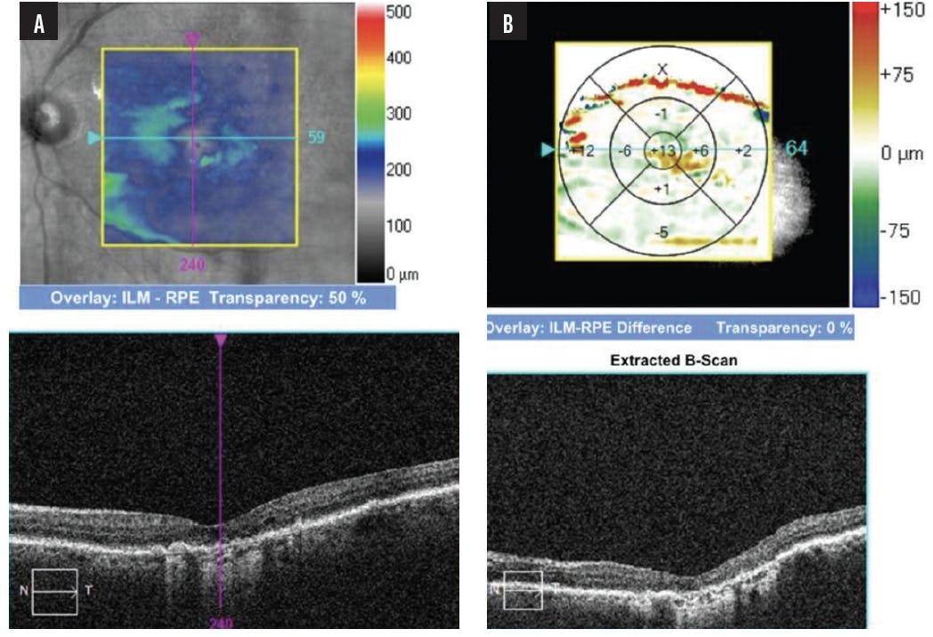
Figure 7. OCT OS depicted central GA with drusen and RPE changes corresponding to fundus photography at presentation (A). At 18 months after her initial visit, GA progression was noted on the ILM-RPE difference map (B, top panel).
Calculations in the ZEISS Advanced RPE Analysis interface compared sub-RPE illumination (signifying GA) within 5 mm of the foveal center and the closest distance to fovea (Figure 8). Significant growth in sub-RPE illumination within 5 mm of the foveal center were noted when comparing her first visit and her most recent visit.

Figure 8. Growth in RPE elevation and sub-RPE illumination within 5 mm of the foveal center were noted when comparing the patient’s first visit with her most recent visit, allowing quantitative characterization of GA progression. Foveal involvement was noted on baseline and remained present in her most recent scan. This assessment was generated by the ZEISS Retina Workplace.
Given the clear progression of GA, this patient elected to begin therapy with pegcetacoplan (Syfovre, Apellis Pharmaceuticals) OS. Longitudinal tracking of lesion growth in this patient will be key to better understanding the effects of therapy. The right eye was monitored given resolution of hemorrhage and fluid with VA limited by disciform scar/RPE tear.