_1710778517.jpg?auto=compress,format)

Marlon Rafael García Roa, MD, of the Instituto Mexicano de Oftalmologia, is the recipient of the 4th Annual NIDEK IMAGE OF THE YEAR Award. His case of a 25-year-old patient with diabetic retinopathy and macular atrophy in the right eye (Figure 1) highlights the imaging capabilities of the Mirante Scanning Laser Ophthalmoscope (NIDEK).

Figure 1. A Mirante 163°* ultra-widefield color image of a right eye with diabetic retinopathy taken from the temporal position when the patient presented in early 2023. The image shows fibrovascular traction in the near periphery, temporally, and superiorly, and a subhyaloid hemorrhage. Laser scars are also visible for 360°.
A 25-year-old female patient presented to our clinic at the Instituto Mexicano de Oftalmología I.A.P. (Santiago de Querétero, Mexico) in early 2023 with diabetic retinopathy in the right eye (Figure 1) and macular atrophy in the left eye (Figure 2). She had undergone surgery 1 year prior for tractional retinal detachment in the left eye, after which the vision was counting fingers at 30 cm.

Figure 2. A Mirante 163°* ultra-widefield color image of the patient’s left eye taken in January 2023 showed a fibrovascular proliferation in the superior far periphery, as well as a remanent of proliferation with perifoveal traction.
My team and I used the Mirante to capture multimodal imaging of the right eye, including ultra-widefield color images and green fundus autofluorescence (FAF). These showed fibrovascular proliferation in the temporal periphery. Also present was a subhyaloid hemorrhage in the posterior pole (Figure 1), and because this configuration increases the risk of acute contraction (the so-called “crunch”) after anti-VEGF injections or laser treatment, these interventions must be used cautiously. Her vision was 20/25 BCVA in the right eye. We decided to treat this eye with vitrectomy with silicone oil, after which her vision stabilized at 20/60 UCVA for 2 to 3 months.
Follow-Up
My team and I saw this patient for a follow-up visit in December 2023. Her postoperative visual acuity in the right eye was 20/40 UCVA and 20/25 BCVA. The eye appeared stable following the vitrectomy (Figures 3–5). We plan to extract the silicone oil in February 2024. The left eye’s acuity remained counting fingers at 30 cm. Using the Mirante OCT, we saw disorganization of the retina’s inner layers and external lines lost (not shown), so we decided not to operate further on this eye. We will closely monitor the patient with Mirante imaging going forward.

Figure 3. A Mirante 163°* ultra-widefield color image of the posterior pole of the patient’s right eye after undergoing vitrectomy showed no fibrovascular traction in the near periphery.

Figure 4. A Mirante 89°* green fundus autofluorescence (FAF) image of the patient’s right eye after undergoing vitrectomy with retained silicone oil reveals points of hypo-fluorescence in the perifoveal area superiorly and inferiorly, indicative of microhemorrhages. Outside the temporal vascular arcades, there are points of hyper-fluorescent spots surrounded by hypofluorescence that indicate areas where laser marks were made during a previous surgery.
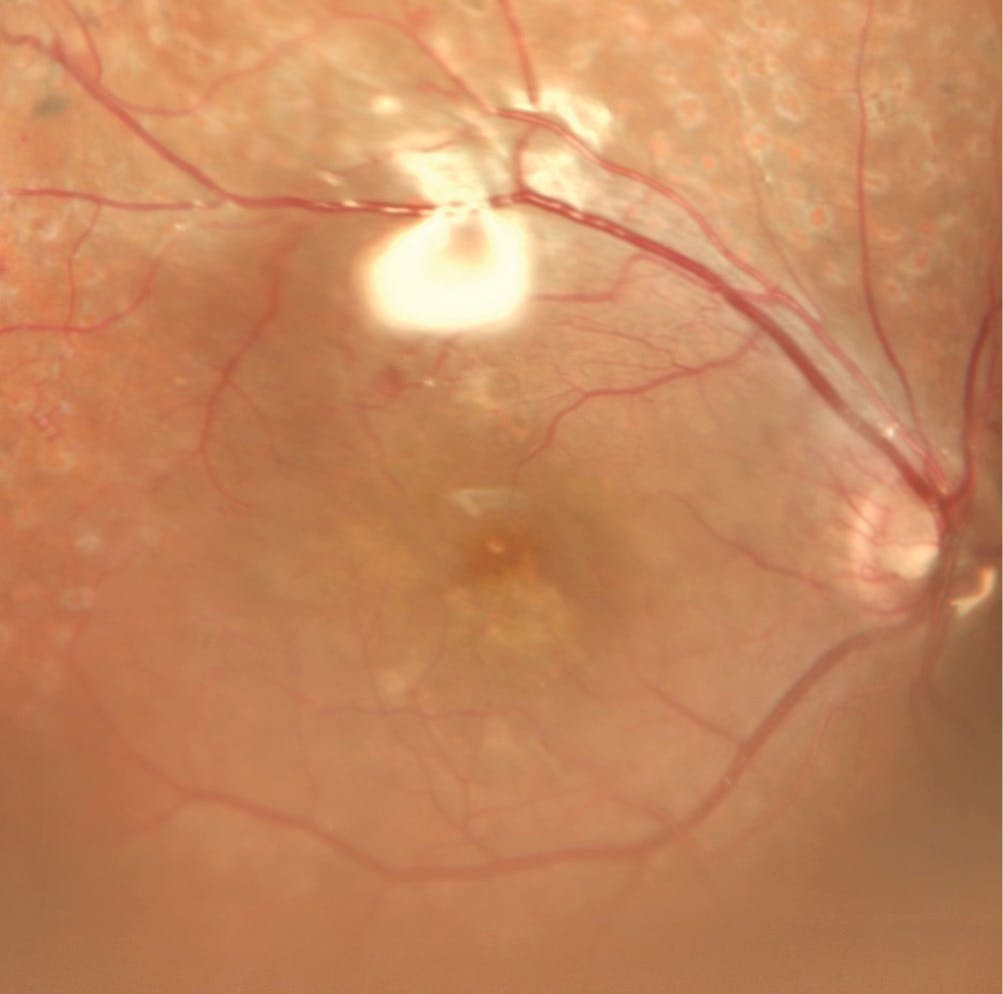
Figure 5. A Mirante 89°* color image of the right eye with the silicone oil evident in the perifoveal area (bright spots). The author and his team will remove the silicone oil at a later visit.
Clinical Benefits of the Mirante
Because there is a high rate of uncontrolled diabetes among the citizens of Mexico, even in young people, diabetic retinopathy is common in our patient population. We receive many referral cases at our institute because of our retina fellowship program. The various imaging modalities on the Mirante have proven very useful in evaluating both adults and children—in particular, these applications help us diagnose retinopathy of prematurity and peripheral retinal diseases. We use the Mirante for diagnosis and treatment decisions, as well as follow-up.
We have had the Mirante device for approximately 2 years; it was our first widefield imaging system. We use it in approximately 50% of our patients, and our most common applications are the green FAF and Retro mode for pathologies such as macular degeneration and central serous retinopathy. We use the color ultra-widefield SLO images for retinal detachments and other traumas. Our patients also appreciate the images the Mirante produces; seeing a visual of a lesion or detachment helps them and their loved ones understand what is happening to their vision.
*Measured from the center of the eye