
One of the best parts of the annual Aspen Retinal Detachment Society (ARDS) meeting—which celebrated 50 years of education in 2022—is the longer format of the sessions and the robust panel discussions. This year, we hosted three top-notch panels to discuss AMD advances and controversies, surgical advances for the complex retina, and the future of the field. I hope you enjoy this recap and consider joining us for the next meeting, which is going to be another fantastic event.
Registration is open for ARDS 2023 (https://aspenretina.com), so mark your calendars for March 4–8, and get ready for more sessions and skiing!
– Timothy G. Murray, MD, MBA
ADVANCES AND CONTROVERSIES IN AMD
The first panel, moderated by Dr. Murray, included Carl C. Awh, MD, FASRS; K. Bailey Freund, MD; Joan W. Miller, MD; and Carl D. Regillo, MD. The group began by discussing the utility of fluorescein angiography (FA) in the era of advanced imaging. “At presentation, if it’s classic wet AMD, with presenting features on OCT, I don’t get a fluorescein angiogram for most patients,” Dr. Awh explained. Although FA is useful for many, if a patient on anti-VEGF therapy has a dry macula after two or three intravitreal injections, no further imaging is needed, he said.
Dr. Freund noted that he often opts for OCT angiography (OCTA) to ensure there is neovascularization before proceeding with therapy. Vitelliform lesions are tough to interpret on FA, and you can get a double-layer sign that’s not really neovascularization, he said. Although a quick audience poll showed that most attendees aren’t using OCTA, Dr. Miller suggested clinicians use it to follow patients after correlating the initial FA imaging with the OCTA imaging.
Next, the panel tackled the tough questions surrounding anti-VEGF therapy. Although everyone turns to anti-VEGF agents as the first-line therapy, the panelists agreed that, often, the choice to start with a branded drug isn’t up to the provider, given some insurance carrier regulations; thus, most start with bevacizumab (Avastin, Genentech/Roche).
Dr. Murray then presented the case of a 90-year-old woman who was unaware of vision loss in the left eye for more than a year and refused any treatment. Her VA was 20/50 OD and 20/200 OS.
“This is really an example of not explaining well to the patient and to the family the implications of not treating that eye, knowing that the fellow eye was at risk,” said Dr. Awh.
Donald J. D’Amico, MD, interjected from the audience with an important patient care pearl: “I challenge the assumption that the patient made a wrong choice here,” he said. “There’s the issue of patient dignity. A difficult part of our training is to do the best we can with those patients, and they made a choice that they’re entitled to make.” Dr. Murray stated that the burden is upon the clinician to accurately portray the benefits and risks. Furthermore, if a patient opts for a non-standard treatment approach, it behooves the clinician to document this well.
Moving on to treatment intervals, the panel debated the existence of true “non-responders” to anti-VEGF therapy. If a patient returns 6 weeks after an injection without improvement, Dr. Murray said he reinjects and brings them back in a few weeks to see if the eye improves in a shorter timeframe.
Dr. Awh cited a study that sampled anterior chamber fluid after a single injection of ranibizumab (Lucentis, Genentech/Roche) and found the drug could last as little as 26 days or as long as 90 days—exactly what we see in the spectrum of patients, he said. “We’re still going to have the frequent flyers.”
SURGICAL ADVANCES IN RETINA
Moderated by Judy E. Kim, MD, the surgical panel included H. Culver Boldt, MD; Steve Charles, MD, FACS, FICS; Dr. D’Amico; Dr. Murray; and Aleksandra V. Rachitskaya, MD (Figure). Dr. Kim began with a query to the panel: what do you think is the most important innovation in retinal surgery?
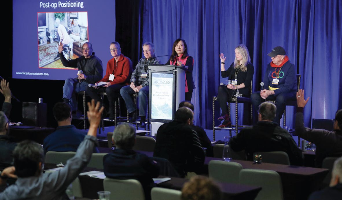
Figure. The surgical panel was lively with friendly banter and audience participation. From left to right: Donald J. D’Amico, MD; Timothy G. Murray, MD, MBA; H. Culver Boldt, MD; Judy E. Kim, MD; Aleksandra V. Rachitskaya, MD; and Steve Charles, MD, FACS, FICS.
Dr. Charles said vitrectomy—and three-port vitrectomy in particular—while Dr. Rachitskaya chose intraoperative OCT and its integration with 3D surgery. Dr. Boldt said the biggest change was the advent of fiber optics, which has allowed the creation of retinal lasers, endoillumination, and more. Dr. D’Amico rounded out the rapid-fire Q&A with the endo laser and the scleral buckle.
Next, Dr. Kim asked about memorable cases the panelists could share with the audience.
Dr. Boldt shared the tale of an 83-year-old patient who fell while in church and hit his head on the pew, leaving him with two fractures and ruptured globes. “He was a 20/25 pseudophakic slipping down the aisle in church, and then suddenly he has no light perception in one eye and bare light perception in the other,” Dr. Boldt said. The initial repair was in no way an elegant solution, and the patient presented to him with “big chunks of the retina removed.” Dr. Boldt put a buckle on him. When talking with the patient’s son, he noticed the son had already lost one eye himself and had a very high prescription in the other. “I asked if he had ever heard of Sticklers,” Dr. Boldt recalled. Turns out, both father and son had Stickler syndrome.
Dr. Murray recalled a case that he and Dr. Boldt handled together as fellows—a case that took close to 14 hours and landed the patient in the intensive care unit after surgery. But the patient “left as one of my happiest patients ever,” Dr. Murray said.
Dr. Charles shared the complicated story of a patient with corneal decompensation, cataract, and an open-funnel retinal detachment.
For Dr. D’Amico, it had to be treating retinal detachments in a convicted psychopathic killer who had been in solitary confinement for years. “They were hideous breaks,” Dr. D’Amico recalled.
Wrapping up the session, Dr. Kim asked for a recap of the current unmet needs in retina. Dr. Rachitskaya is watching the advances in robotics carefully, with the hope that they will help surgeons perform surgeries—such as subretinal gene therapy—with precision.
For Dr. Charles, it’s all about better visualization; Dr. Boldt is hoping for better surgical approaches to hypotony and chronic cystoid macular edema; and Dr. Murray is keeping a close eye on the pharmacotherapy pipeline.
THE FUTURE OF RETINA
The final panel, led by Dr. Miller, included Drs. Charles, Freund, Murray, and Rachitskaya. Dr. Miller started with a discussion of 3D heads-up displays in the OR, which Dr. Rachitskaya said is a wonderful addition for teaching fellows.
The technology also allows for remote surgical education, including live streaming of surgery, said Dr. Miller. Dr. Charles agreed, but also emphasized that the technology does not support remote surgery itself. “You cannot have a lag of even a few milliseconds and operate, so we won’t be operating on patients in India from here,” he explained.
Moving on, Dr. Miller then asked Dr. Freund about the potential advances in screening for retinal diseases, particularly with the use of artificial intelligence and deep learning. “There’s a lot of potential for that with the algorithms that can grade diabetic retinopathy as accurately as retina physicians and the ability to do this in any part of the world,” Dr. Freund said.
Dr. Rachitskaya then discussed the promise of gene therapies. “There is so much unknown in terms of gene therapy, and it is very much disease dependent,” she said. It also depends on the route of delivery, and she hopes suprachoroidal or intravitreal delivery work to keep patients out of the OR.
Dr. Charles played the devil’s advocate by pointing out that new data suggests no visual benefit to gene therapy for inherited retinal diseases. Dr. Rachitskaya countered by saying that, if you’re not losing vision, it may still be a victory.
Dr. Miller then asked about the utility of the port delivery system with ranibizumab (Genentech/Roche). Everyone agreed that the surgery must be done with precision to avoid complications such as conjunctival retraction and erosion and endophthalmitis.
Next, Dr. Miller asked about the pros and cons of telehealth. Dr. Murray expressed his concern that telehealth might not be good for patients with peripheral disease. Still, “it’s going to be an extension of care for patients that we don’t see in our office,” he admitted.
The panel wrapped up with a look at the push for liquid biopsies for diagnosing lymphoma. “If we can get to that, that’s a phenomenal game-changer because having to get tumor tissue is a restriction,” and many will not treat until they have a tissue diagnosis, said Dr. Murray.
Dr. Murray’s last thought was to share that the first treatment for metastatic ocular melanoma, tebentafusp (Immunocore), had been approved in January. “The survival rates are not excellent, but it’s a clear step forward. We are seeing early treatment, and I think we are seeing better outcomes,” he concluded.
With that hope lingering in the air, the panel—and the 50th ARDS meeting—adjourned.


_1784132761.jpg?auto=compress,format&w=75)


